Beyond the Six-Week Clearance — Clinical Pelvic Health for Active Women
Pelvic floor dysfunction is not a normal consequence of childbirth. It is an exceedingly common one — and the distinction matters clinically, because common does not mean inevitable, and it certainly does not mean untreatable. The International Continence Society has described pelvic floor disorders as a "silent epidemic," and that characterization remains apt: a significant portion of the women who present to our clinic with urinary incontinence, pelvic organ prolapse symptoms, pelvic girdle pain, or diastasis recti have been managing these issues for months or years under the assumption that this is simply what happens after having a baby.
It isn't. Or at least — it doesn't have to be. The evidence base for pelvic floor physical therapy in the prenatal and postpartum periods has grown substantially over the past several years. The 2024 Cochrane Review update on pelvic floor muscle training concluded that structured PFMT significantly reduces the risk of urinary incontinence both in late pregnancy and after delivery, with greater benefit observed in women who began training earlier in their pregnancies. A 2025 systematic review and meta-analysis published in the British Journal of Sports Medicine — developed as part of the evidence base for the Canadian Society for Exercise Physiology's postpartum physical activity guidelines — confirmed that pelvic floor muscle training reduces the odds of postpartum urinary incontinence and pelvic organ prolapse with moderate certainty of evidence.
This post outlines what we assess, what the current evidence supports, and what the rehabilitation timeline actually looks like for women from late pregnancy through return to full activity. For a more accessible overview written for athletes at Muók Boxing Georgetown, see our companion piece: Pelvic Floor Health — Before & After Birth.
The Pelvic Floor in Pregnancy — What's Actually Happening
The pelvic floor is a group of muscles, ligaments, and connective tissue forming the base of the pelvis. It supports the bladder, uterus, and rectum; maintains urinary and bowel continence; contributes to sexual function; and plays a load-transfer role in lumbopelvic stability during movement. During pregnancy, this system is under escalating demand that the healthcare system largely does not prepare women for.
Pregnancy-related changes to the pelvic floor begin in the first trimester and compound through delivery. Relaxin — a hormone that peaks in the first trimester and remains elevated through delivery — increases connective tissue laxity throughout the pelvis and lower extremity. This serves an important mechanical purpose during labor, but the same laxity affects joint stability and pelvic floor coordination months before delivery. As the uterus grows and the center of mass shifts anteriorly, lumbar lordosis typically increases, the diaphragm is elevated, and intra-abdominal pressure dynamics change substantially. The load on the pelvic floor muscles increases with every trimester.
The APTA Pelvic Health Division's 2025 evidence summary cites pregnancy-related low back and pelvic girdle pain in 70–86% of pregnant women in the United States. These are the conditions most commonly referred to PT during pregnancy — but they are the downstream presentation of pelvic floor and lumbopelvic dysfunction, not separate entities. Treating the pain without assessing the pelvic floor is managing the symptom without addressing the system.
What prenatal pelvic floor PT actually addresses
A comprehensive prenatal pelvic floor assessment evaluates pelvic floor muscle strength, endurance, and coordination; the presence of hypertonicity (elevated resting tone is more common in athletic women than is widely recognized and is often the cause of pelvic pain rather than weakness); lumbopelvic alignment and load transfer; diaphragmatic and intra-abdominal pressure coordination; and early signs of diastasis recti abdominis — the midline separation of the rectus abdominis that occurs in varying degrees in the majority of pregnancies.
Insufficient resting tone or strength. Associated with stress urinary incontinence, pelvic organ prolapse symptoms, and reduced support during impact loading. The more commonly discussed presentation.
Elevated resting tone or inability to fully relax. Associated with pelvic pain, painful intercourse, perineal trauma during delivery, and — paradoxically — leakage due to poor coordination rather than weakness.
Inter-recti distance widening at the linea alba. Occurs in the majority of pregnancies; clinically significant when associated with poor load transfer, pain, or difficulty generating abdominal wall tension. Not every DRA requires treatment — function matters more than the measurement.
Posterior pelvic girdle pain, sacroiliac joint pain, and pregnancy-related low back pain are distinct diagnoses requiring differentiated assessment. All have strong PT evidence and often respond faster when treatment begins in the prenatal period. See also: our approach to musculoskeletal pain at Root Strength →
"Referral to physical therapy both in the prenatal and postnatal period is currently not considered standard of care — despite robust evidence that early intervention reduces the prevalence of musculoskeletal pain, diastasis recti, and pelvic floor dysfunction." — International Journal of Sports Physical Therapy, 2022
The Delivery Variable — What Changes Based on Mode of Birth
Pelvic floor sequelae differ meaningfully between vaginal and cesarean delivery — but cesarean section does not protect the pelvic floor from dysfunction, and this is one of the most persistent misconceptions we encounter clinically.
Vaginal delivery
Vaginal delivery, particularly with prolonged second stage, instrumental delivery (forceps or vacuum), and significant perineal tearing, is associated with greater immediate pelvic floor muscle trauma, levator ani injury, and pudendal neuropathy. Obstetric anal sphincter injuries (OASIS) — third and fourth-degree tears — occur in roughly 1–5% of vaginal deliveries and require specific postpartum assessment and rehabilitation. The research on prenatal perineal massage — initiated from 34–36 weeks — shows reduced rates of perineal trauma and perineal pain postpartum, though it does not affect episiotomy rates. This is an area where prenatal PT provides direct, evidence-supported benefit.
Cesarean section
Women who deliver by cesarean section still experience nine months of pelvic floor loading during pregnancy, hormonal ligament laxity, and the full range of prenatal pelvic floor changes — they simply avoid the acute delivery trauma. However, cesarean delivery introduces a surgical scar — the lower uterine segment incision and the layers of abdominal fascial repair — that frequently causes adhesion, restricted mobility, and dysesthetic pain if not treated. C-section scar mobilization is a standard component of our postpartum PT assessment, typically initiated at 6–8 weeks post-delivery once the incision has closed, and the literature supports that early scar treatment reduces long-term mobility restriction and pain.
We assess C-section scars as a standard part of postpartum evaluation regardless of whether the patient mentions scar-related symptoms. Restricted scar mobility can contribute to bladder dysfunction, hip flexor inhibition, lumbopelvic pain, and altered movement mechanics — none of which feel obviously related to the incision site. If you delivered by cesarean and have never had your scar assessed, that assessment is worth having.
The Evidence on Pelvic Floor Muscle Training
Pelvic floor muscle training (PFMT) — structured, progressive exercise targeting the pelvic floor muscles — is the most evidence-supported conservative intervention for both prevention and treatment of pelvic floor dysfunction across the prenatal and postpartum periods. The evidence on what PFMT actually does, and at what dose, has become substantially more precise.
Prenatal PFMT — prevention and preparation
A 2024 systematic review and meta-analysis published in Acta Obstetricia et Gynecologica Scandinavica evaluated the effect of PFMT during pregnancy on urinary incontinence, episiotomy rates, and perineal tearing. The analysis found that PFMT begun in early pregnancy — before symptoms develop — reduces the incidence of urinary incontinence in late pregnancy. A separate 2024 systematic review in Neurourology and Urodynamics found that aerobic and resistance exercise combined with PFMT during prenatal care was effective for both prevention and treatment of urinary incontinence. The evidence for starting PFMT earlier in pregnancy is consistently stronger than for initiating it in the third trimester or postpartum.
Equally important: the research supports a significant reduction in the duration of the second stage of labor in women who performed structured PFMT during pregnancy. A pelvic floor that has been trained to contract, coordinate, and — critically — fully relax, is better positioned to facilitate fetal descent than a floor that is either weak or hypertonic.
Postpartum PFMT — treatment and rehabilitation
The 2025 meta-analysis in the British Journal of Sports Medicine, conducted as part of the evidence base for the Canadian Society for Exercise Physiology's postpartum guidelines, analyzed data from seven randomized controlled trials involving 1,930 participants and found that PFMT reduced the odds of postpartum urinary incontinence by 37%. Pelvic organ prolapse odds were similarly reduced. These findings carry moderate certainty of evidence — which, in a clinical context involving a non-pharmacological intervention with no meaningful adverse event profile, represents a strong basis for clinical recommendation.
The practical implication: PFMT is first-line treatment for postpartum urinary incontinence and pelvic organ prolapse. It is not a supplement to other treatment — it is the treatment. The question is not whether to do it but how to program it correctly for an individual patient's presentation, which varies considerably and is why a PT assessment informs the program rather than a generic exercise sheet.
Our PT team provides comprehensive prenatal and postpartum pelvic floor assessment and rehabilitation. Internal and external assessment available. No referral required in Washington State. Learn about our PT department →
The Postpartum Return-to-Activity Framework
The standard postpartum clearance model — a single 6-week OB/GYN visit followed by a blanket "cleared for activity" — is not functionally adequate for most women and is particularly insufficient for athletes or active individuals. The assessment at 6 weeks does not include pelvic floor muscle evaluation, load transfer testing, or movement screen. A woman can receive clearance at 6 weeks while having clinically significant diastasis recti, a hypertonic pelvic floor, and an incompletely healed perineal repair — all of which will be exacerbated by returning to running or lifting without appropriate progression.
The following framework is adapted from the evidence in postpartum rehabilitation literature, including the Maximizing Recovery in the Postpartum Period commentary published in the International Journal of Sports Physical Therapy and the return-to-running framework from the postpartum elite athlete literature (PMC, 2025).
- Pelvic floor muscle awareness and gentle activation — not strengthening. The goal is neuromotor reconnection, particularly after epidural or significant perineal trauma that may cause temporary proprioceptive impairment
- Diaphragmatic breathing coordinated with pelvic floor — foundational for all subsequent rehabilitation
- Light walking as tolerated; perineal care following vaginal delivery or incision care following cesarean
- No abdominal loading, no impact, no resistance training
- Edema management and positioning as indicated
- Structured PFMT progressing from endurance holds (10-second contractions) to quick-flick recruitment (10 rapid contractions) — both domains are necessary for complete continence function
- Postural correction and lumbopelvic alignment — anterior pelvic tilt and ribcage flare are common postpartum postures that compromise pelvic floor mechanics
- Diastasis recti assessment and management — coning or doming with any loaded exercise indicates inadequate linea alba tension management; those movements should be modified until control is established
- Gentle lower body loading: bridge progressions, clamshells, lateral band walks — coordinated with breath and pelvic floor
- C-section scar mobilization beginning at 6–8 weeks, once the incision has closed
- Walking progression — duration and pace increasing as tolerated without symptom provocation
- Resistance training reintroduction — squats, deadlifts, pressing — at reduced load from pre-pregnancy baseline, with attention to intra-abdominal pressure management and pelvic floor response
- Anti-rotation and anti-extension core progressions replacing early-postpartum stability work
- Hip and gluteal strengthening — the hip abductors and external rotators are critical load-transfer partners for the pelvic floor and are frequently de-conditioned postpartum
- Functional movement patterns under load: carries, step-ups, single-leg progressions. The principles here mirror those we apply in any return-to-training after injury — graduated load, monitored response
- No running, jumping, or impact activities until pelvic floor can manage the load criteria below
- Run/walk intervals progressing to continuous running — the specific progression timeline depends on Phase 3 readiness, not gestational age alone
- Impact monitoring: urinary leakage, pelvic heaviness or pressure, lumbopelvic pain, and pelvic floor fatigue are all signals to reduce load and reassess
- Sport-specific loading reintroduced after foundational running tolerance is established
- Athletes returning to high-impact, high-intensity activity — including Muay Thai at Muók Boxing, weightlifting, and CrossFit — require individualized progression. The 12-week threshold is a floor, not a clearance date. For Muay Thai athletes specifically, return to sparring involves criteria beyond running tolerance; see our companion guide for that full framework
- Nutritional considerations: bone density, RED-S risk, and energy availability are particularly relevant for breastfeeding athletes returning to high-volume training
What We Assess at Root Strength
A pelvic floor assessment at Root Strength is not a questionnaire and a printed Kegel instruction sheet. Our Doctors of Physical Therapy conduct a full clinical evaluation that includes external and, where clinically appropriate and consented, internal assessment of pelvic floor muscle function. The internal assessment — where the clinician evaluates muscle tone, strength, coordination, and tender points through vaginal or rectal examination — is the only way to definitively characterize whether a pelvic floor is hypotonic, hypertonic, or coordinating correctly. Many women who have been told their pelvic floor is "weak" have never had an internal assessment and have been programmed to do Kegel exercises — which are contraindicated in hypertonicity and will worsen the presentation.
Pelvic health assessments at Root Strength are led by Dr. Lorrainne, a Doctor of Physical Therapy currently completing an orthopedic residency in pursuit of board-certification in orthopedic physical therapy. Lorrainne has extensive experience working with women navigating pelvic floor dysfunction across the prenatal and postpartum spectrum — and a particular focus on active women and athletes who want to return to training after having a baby. If you've been hesitant to book because you weren't sure who would be seeing you: it's Lorrainne, and she's the right fit for exactly this kind of care. Book with Lorrainne →
Prenatal assessment
We recommend a prenatal pelvic floor assessment for any woman who is planning to continue or resume physical training during pregnancy, who has a history of pelvic floor symptoms, who is preparing for delivery and wants to optimize the mechanical conditions for labor, or who has any of the musculoskeletal conditions associated with pregnancy (pelvic girdle pain, pubic symphysis dysfunction, round ligament pain). Ideally, this occurs in the second trimester — early enough that training has time to be meaningful before delivery. Book a prenatal assessment →
Postpartum assessment
We recommend a postpartum pelvic floor assessment for all women at 6–8 weeks following delivery, regardless of symptom status. Asymptomatic does not mean the pelvic floor has recovered — it means symptoms haven't yet been provoked. Many women present to us at 3–6 months postpartum with incontinence that developed after returning to running that had been symptom-free in the early postpartum period. Earlier assessment and earlier appropriate loading prevents that outcome. Our PT department accepts most major insurance and requires no physician referral in Washington State.
The following symptoms at any point during pregnancy or postpartum require clinical evaluation before returning to exercise: any urinary or fecal leakage; a sensation of heaviness, pressure, or bulging in the vagina (possible prolapse); pelvic pain that affects daily activities or sexual function; ongoing perineal or scar-related pain beyond 8 weeks; or any incontinence that persists beyond 12 weeks postpartum despite exercise. Washington State has direct access to PT — no physician referral is required to book an assessment.
Pelvic Floor Health and the Athletic Postpartum Patient
The postpartum elite athlete literature — including a 2025 PMC review on return to running for postpartum elite and sub-elite athletes — is consistent on one point: current evidence is insufficient to guide highly active women through postpartum return, and the absence of guidance has led to inconsistent protocols, unnecessary delays, and preventable injury. The same gaps exist in the recreational athlete population, who receive even less individualized guidance.
At Root Strength, a meaningful portion of our postpartum PT patients are active women — members of Muók Boxing, recreational runners, CrossFit athletes, and gym-based strength trainees — who want to return to training and lack a clear, clinically grounded framework for doing so. The return-to-sport timeline for these patients is not determined by gestational age or a 6-week OB clearance. It is determined by objective clinical criteria: pelvic floor function under load, lumbopelvic stability, diastasis recti management, and the absence of provoked symptoms during graduated activity progression. The principles are the same whether someone is returning to Muay Thai for fitness or competitive sparring.
We work directly with the coaching team at Muók Boxing to ensure that members who are pre- or postpartum have a training plan that is safe and appropriately progressive for where they are clinically. That coordination — between the PT assessment and the coaching environment — is what makes the difference between a safe return and one that creates a setback. Our approach to postpartum return mirrors the framework we apply to concussion return-to-sport and injury rehabilitation broadly: criteria-driven, not calendar-driven.
Ready for a Pelvic Health Assessment?
Dr. Lorrainne sees patients on-site at Root Strength Georgetown. No referral required. Most major insurance accepted. She'll evaluate your pelvic floor function, screen for diastasis and lumbopelvic dysfunction, and build a return-to-activity plan built around where you are right now — and where you want to get back to.
Book with Lorrainne →- Beamish NF, Davenport MH, Ali MU, et al. Impact of postpartum exercise on pelvic floor disorders and diastasis recti abdominis: a systematic review and meta-analysis. British Journal of Sports Medicine. 2025;59(8):562–575. doi:10.1136/bjsports-2024-108619
- Woodley S, Dumoulin C. Pelvic floor muscle training for preventing and treating urinary incontinence during pregnancy and after childbirth: A Cochrane Review. Cochrane Database of Systematic Reviews. 2024;(1):CD012279.
- Zhang R, et al. Influence of pelvic floor muscle training alone or as part of a general physical activity program during pregnancy on urinary incontinence, episiotomy and third- or fourth-degree perineal tear: Systematic review and meta-analysis. Acta Obstetricia et Gynecologica Scandinavica. 2024;103:1015–1027. doi:10.1111/aogs.14744
- Stephenson R, Cathcart D. Physical Therapy Examination, Evaluation, and Treatment of Musculoskeletal Disorders during Pregnancy and Postpartum. In: The Physical Therapist's Guide. Routledge: New York. 2025.
- Deering RE, Donnelly GM, Brockwell E, et al. Return to running postpartum: updated guidelines for medical professionals. British Journal of Sports Medicine. 2025. doi:10.1136/bjsports-2024-109104
- Donnelly GM, Moore IS, Brockwell E, et al. Reframing return-to-sport postpartum: the 6 Rs framework. British Journal of Sports Medicine. 2022;56(5):244–245.
- Maximizing recovery in the postpartum period: a timeline for rehabilitation from pregnancy through return to sport. International Journal of Sports Physical Therapy. 2022. doi:10.26603/001c.37863
- Return to running for postpartum elite and subelite athletes. PMC. Published 2025 May–Jun. PMID: 11569573
- Santos AC, et al. Effectiveness of group aerobic and/or resistance exercise programs associated with pelvic floor muscle training during prenatal care for the prevention and treatment of urinary incontinence. Neurourology and Urodynamics. 2024;43(1):205–218. doi:10.1002/nau.25309
- APTA Pelvic Health Division. Pregnancy & Postpartum Physical Therapy — Evidence Highlight. 2025. aptapelvichealth.org
Concussion — Assessment, Management & Return to Sport

Concussion management has undergone more significant evidence-based revision in the past five years than in the preceding two decades. The publication of the 6th International Consensus Statement on Concussion in Sport — the Amsterdam Statement — in the British Journal of Sports Medicine in June 2023, following the October 2022 conference, represents the current gold standard for concussion identification, assessment, and management. Several of its recommendations represent meaningful departures from earlier guidelines that remain in widespread clinical use.
This post summarizes the current state of the evidence as it applies to our patient population at Root Strength — primarily active individuals and combat sports athletes — and outlines the clinical framework our PT team applies when assessing and managing concussive injury.
Definition and Pathophysiology
The Amsterdam 2022 Consensus Statement defines sport-related concussion (SRC) as a traumatic brain injury caused by a direct blow to the head, neck, or body, in which an impulsive force is transmitted to the brain. The injury results in a range of clinical symptoms and signs that may or may not involve loss of consciousness, and which cannot be explained by drug, alcohol, or medication use, other injuries, or comorbid conditions.
Critically, concussion does not demonstrate abnormalities on standard structural neuroimaging — MRI and CT are typically normal. This is not because the injury is minor; it reflects the nature of the pathophysiology. Concussion produces a neurometabolic cascade: ionic flux across neuronal membranes, release of excitatory neurotransmitters, impaired mitochondrial oxidative metabolism, reduced cerebral blood flow, and axonal dysfunction. The brain's energy demand increases at the same time its capacity to generate energy is compromised — a cellular energy crisis that manifests as the clinical syndrome we recognize as concussion.
This metabolic vulnerability is why the period immediately following concussion carries elevated risk. A second impact during this window — before neurometabolic recovery is complete — can produce disproportionately severe consequences. Second-impact syndrome, while relatively rare, is associated with rapid catastrophic cerebral edema and remains a serious concern in return-to-sport decision-making.
Clinical Presentation — Symptom Domains
Concussion symptoms span four domains. A thorough assessment addresses all four, not just the most visible physical symptoms.
Headache (most common), pressure in the head, nausea, vomiting, visual disturbance, photosensitivity, phonosensitivity, balance disturbance, dizziness, fatigue, sleep disruption.
Feeling "in a fog," slowed processing speed, difficulty concentrating, memory impairment (particularly anterograde), difficulty with word-finding, reduced academic or occupational performance.
Irritability, emotional lability, anxiety, low frustration tolerance, depression. Often underreported, particularly in competitive athletes. The Amsterdam Statement emphasizes the importance of mental health assessment as part of the concussion evaluation.
Hypersomnia (sleeping more than usual), hyposomnia (difficulty sleeping), or disrupted sleep architecture. Sleep disturbance both reflects and compounds other concussion symptoms, and often requires specific management.
The following presentations require emergency evaluation before any further assessment: loss of consciousness (any duration), seizure or tonic posturing, repeated vomiting, progressively worsening headache, focal neurological deficit (weakness, numbness, double vision, slurred speech), deteriorating conscious state, or Glasgow Coma Scale below 15. These presentations may indicate intracranial hemorrhage or other structural injury requiring imaging and emergency intervention.
Assessment Tools — Current Best Practice
The Amsterdam 2022 Statement introduced updated versions of the primary concussion assessment tools. These replace earlier versions and are now the recommended standard.
Sideline assessment — CRT6 and SCAT6
The Concussion Recognition Tool 6 (CRT6) is designed for non-medical personnel — coaches, parents, and athletes themselves — to identify when a concussion may have occurred. The Sport Concussion Assessment Tool 6 (SCAT6) is the current clinical sideline tool for healthcare providers, including elements of symptom assessment, cognitive evaluation, and balance testing. Both tools are most effective within the first 72 hours post-injury. The SCAT6 and CRT6 replace their previous versions and should not be used interchangeably with SCAT5 or older iterations.
Subacute office assessment — SCOAT6
The Sport Concussion Office Assessment Tool 6 (SCOAT6) — a new tool introduced at Amsterdam 2022 — is designed for the clinical follow-up assessment beyond 72 hours. It incorporates multimodal evaluation: symptom scales, balance measures, cognitive testing, oculomotor and vestibular assessment, mental health screening, and sleep assessment. This comprehensive approach reflects the growing recognition that concussion is not a single-domain injury and that adequate assessment requires evaluating all affected systems.
What we assess at Root Strength
Our PT team's concussion assessment protocol includes symptom inventory across all four domains, cervical spine assessment (cervicogenic headache and neck pain frequently co-occur with SRC and require differentiation), vestibular and oculomotor screening, balance and postural control evaluation, and a graded exertion test where clinically appropriate. We work collaboratively with referring physicians and, for athletes, with their coaching staff at Muók Boxing to ensure assessment findings directly inform the return-to-sport timeline.
"Strict rest until complete symptom resolution is no longer supported by the evidence. The current literature supports early sub-threshold aerobic exercise as both safe and beneficial for recovery when properly supervised." — Amsterdam Consensus Statement, 2023
Management — What the Current Evidence Supports
The most clinically significant update in the Amsterdam 2022 Statement concerns rest recommendations. Earlier guidelines universally recommended complete physical and cognitive rest until all symptoms resolved. The evidence has shifted substantially from this position.
The relative rest period — 24 to 48 hours
A brief period of relative rest — reduced physical and cognitive demands without complete isolation — is still recommended immediately post-injury. This is not indefinite. After 24–48 hours, the evidence supports gradual reintroduction of light activity rather than continued rest.
Early sub-threshold aerobic exercise
Research published in JAMA Pediatrics demonstrated that individualized sub-threshold aerobic exercise — activity calibrated to stay below the heart rate threshold that exacerbates symptoms — prescribed as early as two days after SRC safely accelerated recovery and reduced the incidence of persistent post-concussion symptoms compared to strict rest. A systematic review and meta-analysis published in the British Journal of Sports Medicine (Leddy et al., 2023) confirmed these findings across multiple populations.
The clinical implication: the post-concussion period is not a period of complete inactivity for most patients. It is a period of carefully managed, symptom-guided progressive activity. The threshold concept is central — any activity that provokes symptoms is too much; activity that does not provoke symptoms is generally appropriate and often beneficial.
Cervicovestibular rehabilitation
The Amsterdam Statement introduced a new recommendation for cervicovestibular rehabilitation for athletes presenting with neck pain, headache, dizziness, or balance disturbance following SRC. Evidence supports that cervical and vestibular dysfunction frequently co-occur with concussion and contribute significantly to symptom burden and prolonged recovery. Early vestibular rehabilitation — initiated within the first week post-injury — has been associated with faster recovery in athletes compared to delayed initiation. This is an area where physical therapy has a direct, evidence-supported role in concussion management.
Our PT team provides cervicovestibular assessment and rehabilitation as part of our concussion management protocol — one of the most evidence-supported interventions for reducing recovery time. Learn about our PT department →
The 6-Step Return-to-Sport Protocol — Amsterdam 2023
The structured return-to-sport (RTS) protocol from the Amsterdam Consensus Statement is the current evidence-based framework for progression from rest to full athletic participation. Each step requires a minimum of 24 hours. Athletes experiencing concussion-related symptoms during Steps 4–6 should return to Step 3 and re-establish full symptom resolution before progressing. Written medical clearance is required before Step 5.
- Normal activities of daily living permitted — light walking, basic self-care
- Reduced screen time, reduced academic or occupational cognitive load
- No sport participation of any kind
- This phase ends at 24–48 hours, not upon symptom resolution — a critical departure from older guidelines
- Stationary cycling, light walking, or easy swimming
- No resistance training, no body contact, no head movement risk
- Amsterdam 2022 introduced the sub-categorization of Step 2 into 2A (light, ≤55% HR max) and 2B (moderate, ~70% HR max) — a refinement of the earlier single aerobic step
- Mild symptom exacerbation (<2 points on 0–10 scale, resolving within 1 hour) is acceptable; significant or prolonged exacerbation is not
- Moderate jogging, brief running, moderate-intensity stationary cycling
- Moderate-intensity weightlifting at reduced load from baseline
- Body and head movement introduced but no contact risk
- Symptom monitoring throughout and for 24 hours following each session
- Running drills, agility work, sport-specific conditioning
- For Muay Thai athletes: shadow boxing, heavy bag work, footwork drills — no partner contact
- Full intensity of non-contact sport-specific training
- Cervicovestibular rehabilitation continues in parallel if indicated
- Pad work, partner drilling at controlled intensity, clinch technique with cooperative partners
- Full training intensity on non-contact elements of the sport
- Amsterdam 2022 introduced an explicit medical clearance requirement before Step 4 — this step requires clinician assessment confirming symptom resolution with exertion
- Cognitive function and other clinical findings must also have normalized — not just symptom absence
- Full sparring at graduated intensity — light technical contact before full intensity
- Requires written determination of readiness from a healthcare provider
- Ongoing symptom monitoring — any return of concussion symptoms requires return to Step 3
- For Muay Thai athletes, this includes return to clinch sparring and checked kicks — monitoring for symptom provocation with each session
- Unrestricted return to all sport activities including competition
- Maintained symptom-free status with all levels of exertion
- Neuromuscular training and neck strengthening maintained as ongoing prevention measures
- Athletes with a history of multiple concussions: individualized discussion regarding retirement from contact sport, as recommended by the Amsterdam Statement
Persistent Post-Concussion Symptoms
Most adults recover from sport-related concussion within 10–14 days. When symptoms persist beyond four weeks, the term persistent post-concussion symptoms (PPCS) is applied. The Amsterdam 2022 Statement notes that PPCS should be approached through a multidisciplinary lens — the symptoms frequently have multiple contributing drivers rather than a single pathophysiological cause.
The most common drivers of PPCS identified in current research include vestibular and visual dysfunction, cervical musculoskeletal dysfunction, autonomic dysregulation (particularly exercise intolerance), sleep disturbance, and psychological factors including anxiety and depression. Each of these requires specific targeted management rather than continued general rest.
For patients presenting with PPCS, our PT team conducts a systematic assessment to identify the primary drivers and address them directly — vestibular rehabilitation, cervical treatment, graded aerobic exercise protocols, and coordination with mental health providers where indicated. This is one of the most clear clinical applications of physical therapy in concussion management, supported by the Amsterdam 2022 recommendations.
Long-Term Risk and the CTE Question
Concerns about the long-term neurological consequences of repeated head trauma — including chronic traumatic encephalopathy (CTE) — are legitimate and deserve an honest clinical discussion. The Amsterdam 2022 Statement acknowledges that current evidence is insufficient to establish a definitive causal relationship between sport-related concussion and CTE at the population level. CTE remains a post-mortem neuropathological diagnosis and cannot be clinically diagnosed in living individuals.
What the evidence does support is that proper management of individual concussions — immediate removal, structured graduated return, no return while symptomatic — meaningfully reduces cumulative head trauma exposure. Research published in the British Journal of Sports Medicine (Eliason et al., 2023) demonstrated that structured concussion management programs were associated with a 63% reduction in recurrent concussion rates. The individual concussion that is well-managed represents a fundamentally different exposure than the same injury trained through.
For athletes training at Muók Boxing or in our Root Strength programs, this is the practical takeaway: how you manage each concussion is the variable within your control. The cumulative risk reduction from consistent proper management over a training career is substantial.
Need a Concussion Assessment?
Our Doctors of Physical Therapy conduct concussion assessments on-site at Root Strength Georgetown. No referral required. Most major insurance accepted. We'll evaluate your symptoms, assess vestibular and cervical function, and build a return-to-sport plan grounded in the current evidence.
Book an Assessment →- Patricios JS, Schneider KJ, Dvorak J, et al. Consensus statement on concussion in sport: the 6th International Conference on Concussion in Sport–Amsterdam, October 2022. British Journal of Sports Medicine. 2023;57(11):695–711. doi:10.1136/bjsports-2023-106898
- Broglio SP, Harezlak J, Rowson S, et al. Bridge statement: management of sport-related concussion. Journal of Athletic Training. 2024;59(3):225–242. doi:10.4085/1062-6050-0046.22
- Leddy JJ, Burma JS, Toomey CM, et al. Rest and exercise early after sport-related concussion: a systematic review and meta-analysis. British Journal of Sports Medicine. 2023;57(11):762–770.
- Leddy JJ, Mannix R, Willer B, et al. Early subthreshold aerobic exercise for sport-related concussion: a randomized clinical trial. JAMA Pediatrics. 2019;173(4):319–325. doi:10.1001/jamapediatrics.2018.4397
- Anderson M, et al. Early vestibular rehabilitation initiation is associated with faster recovery after sport-related concussion. Journal of Science and Medicine in Sport. 2025;28(3):222–227.
- Eliason PH, Galarneau JM, Kolstad AT, et al. Prevention strategies and modifiable risk factors for sport-related concussions and head impacts: a systematic review and meta-analysis. British Journal of Sports Medicine. 2023;57(12):749–761.
- Hallaçeli H, Davut S, Özbek A, et al. Epidemiological analysis of athlete injuries in Muay Thai in-ring matches. Injury Epidemiology. 2025;12:28. doi:10.1186/s40621-025-00569-x
- Doherty CS, Barley OR, Fortington LV. Incidence of health problems in Australian MMA and Muay Thai competitors: a 14-month study. Sports Medicine – Open. 2025;11:60. doi:10.1186/s40798-025-00880-3
- Kaguturu N, et al. Advancing sports-related concussion management: evidence-based protocols and emerging diagnostics. Journal of Integrated Primary Care. 2025;2(1):Art.3.
- CDC HEADS UP. Returning to sports. Updated September 15, 2025. Centers for Disease Control and Prevention. cdc.gov/heads-up
Shoulder Pain — Why It Happens, What It Means, and How to Get Rid of It
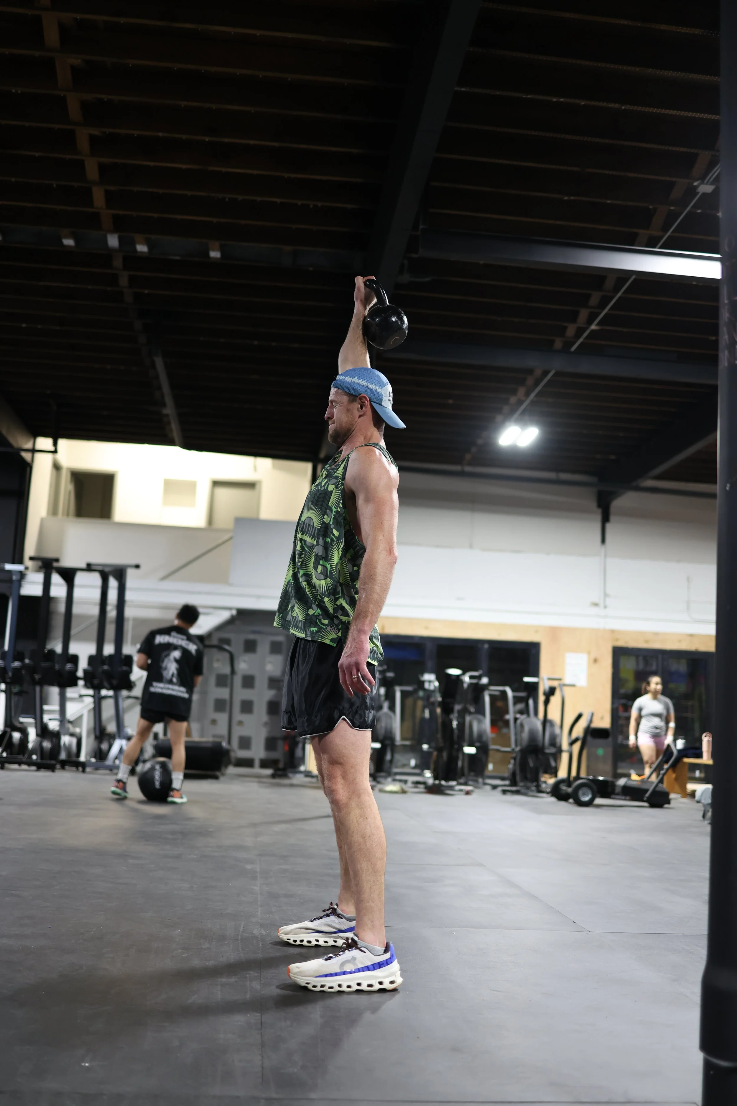
Shoulder pain has a way of making everything harder. Reaching for something on a high shelf. Putting on a jacket. Rolling over in bed. Lifting anything overhead at the gym. It shows up in the most ordinary moments, and once it's there, it's hard to ignore.
It's also one of the most common things we treat at Root Strength. We see it in people who lifted too heavy too fast, in people who spend all day at a desk with their shoulders rounded forward, in people who slept on it wrong and woke up unable to raise their arm, and in people who genuinely have no idea when or how it started — just that it's been bothering them for weeks and they've been hoping it would go away on its own.
Sometimes it does. Often it doesn't. And the longer you wait, the more compensation patterns develop around it — and those take longer to undo than the original problem.
Here's what we see most often, why it happens, and what the path back to a pain-free shoulder actually looks like.
Shoulder pain rarely comes from one dramatic moment. Most of the time it builds quietly — from repetitive load, poor positioning, or simply asking a joint to do more than the muscles around it are prepared to support.
Where Shoulder Pain Usually Comes From
The shoulder is the most mobile joint in the body — it can move in more directions than any other joint you have. That mobility comes at the cost of stability. The shoulder relies almost entirely on the surrounding muscles to keep it in place and moving correctly, which means when those muscles are weak, fatigued, or imbalanced, the joint starts taking load in ways it wasn't designed to handle.
Weight training — the most common culprit we see
Bench press, overhead press, lateral raises, pull-ups — all of these are excellent exercises, and all of them have the potential to irritate the shoulder when volume increases too fast, technique breaks down under fatigue, or the muscles responsible for stabilizing the joint aren't strong enough to support the load being asked of them. The rotator cuff — four small muscles that hold the ball of the shoulder in its socket — is almost universally undertrained relative to the larger pushing and pulling muscles most people focus on. That imbalance is the underlying driver of the majority of gym-related shoulder problems we see.
Desk work and posture
Sitting at a computer for eight hours with your shoulders rounded forward doesn't feel like much in the moment. Over months and years, it changes the resting position of the shoulder blade, tightens the front of the shoulder, and weakens the muscles at the back. When you then go to the gym and push or pull overhead, the joint isn't starting from a neutral position — it's already compromised. This is why so many people who "don't lift that heavy" still end up with shoulder pain. The problem isn't what they're doing in the gym. It's what eight hours of desk posture did to the shoulder before they got there.
Repetitive overhead work
Painters, electricians, construction workers, people who stock shelves — any job that involves repeated overhead reaching places sustained demand on the rotator cuff and the structures around it. Unlike a single heavy lift, the problem here is volume over time. The tissue doesn't get injured in one session; it accumulates stress session after session until it crosses a threshold and starts to complain.
Sleeping position
Sleeping on the same shoulder every night compresses the joint for hours at a time. For a shoulder that's already irritated or vulnerable, this is often what tips it over the edge from "occasionally sore" to "can't raise my arm without pain." Waking up with a stiff or painful shoulder after sleeping on it is a signal the joint isn't tolerating that load well — not something to push through.
Doing nothing — the underrated cause
Sedentary shoulders get weak. Weak shoulders get injured more easily. A large proportion of the shoulder problems we see aren't from overuse — they're from a shoulder that hasn't been asked to do enough for long enough, and then suddenly is. Starting a new workout program, picking up a new sport, or just moving into a house and doing a weekend of heavy lifting are all common triggers when the shoulder musculature hasn't been maintained.
The Most Common Diagnoses — What They Mean in Plain Language
If you've seen a doctor for shoulder pain, you may have heard one of these terms. Here's what they actually mean and what they feel like.
The most common shoulder diagnosis. Pain on the outside or front of the shoulder when you raise your arm — especially between 60 and 120 degrees of elevation. Often feels like a pinch or catch. Worse after overhead lifting or pressing. Usually develops from rotator cuff weakness and poor scapular positioning.
Irritation or degeneration of one or more of the rotator cuff tendons — typically the supraspinatus. Aches during and after lifting. Often worse the morning after a heavy session. Develops when load increases faster than the tendon can adapt. Responds very well to progressive loading when managed correctly.
Pain at the very top of the shoulder where the collarbone meets the shoulder blade. Tender to direct pressure. Aggravated by reaching across your body and by heavy pressing. Common in people who bench press frequently without balancing with posterior shoulder work.
Progressive stiffness and pain that limits motion in all directions — not just one. Often develops after a period of immobility or following an injury that wasn't mobilized properly. Recovers fully in most cases but takes longer than other shoulder conditions and requires a specific approach.
If your shoulder pain wakes you from sleep, if you have significant weakness without proportional pain, if pain followed a specific traumatic incident, or if it has not improved at all after two weeks of reduced load — these are signals that warrant an assessment before you continue loading the joint. Come in and let us take a look before it becomes a longer problem.
Why Rest Alone Usually Doesn't Fix It
The instinct when something hurts is to rest it and wait for it to heal. For some injuries, that's the right call. For most shoulder problems, it's not — and here's why.
The majority of shoulder pain develops because the muscles supporting the joint aren't strong enough or positioned correctly enough to handle the demands being placed on them. Rest reduces the load temporarily, which reduces the pain. But it doesn't build the strength that was missing. When you return to the same activities — lifting, working, sleeping on it — the same weakness is still there, and the problem returns.
The athletes and patients who resolve shoulder pain quickly and keep it from coming back are the ones who use the period of reduced load to build what was missing in the first place. That's the core of what good shoulder rehabilitation looks like.
The 4-Phase Rehab Approach Our PT Team Uses
- Identify and temporarily remove the specific activities driving the pain — overhead pressing, heavy bench, repetitive reaching — without stopping all activity
- Modify your gym routine around the shoulder: lower body training, core work, and cardio can all continue unmodified
- If you work a desk job, address workstation positioning — monitor height, arm support, chair setup — because this is often contributing more than people realize
- Begin gentle range of motion work: pendulum swings, wall slides, and supported elevation to maintain mobility while reducing active load
- Ice for acute flares (first 48–72 hours of a new flare): 15–20 minutes on the painful area, cloth between ice and skin
- Rotator cuff strengthening: side-lying external rotation, banded external rotation, internal rotation with band — these are the exercises that address the most common deficit driving shoulder pain
- Scapular stabilization: prone Y-T-W raises, face pulls, band pull-aparts — essential for restoring the shoulder blade positioning that creates room for the rotator cuff to function without impingement
- Posterior shoulder stretching: cross-body stretch and sleeper stretch to address the tightness that develops from sustained desk posture and pressing-heavy training programs
- Postural retraining: if desk work is a contributing factor, specific exercises to strengthen the mid-back and retrain resting shoulder position
- Begin returning to gym work with modified loading: lighter weight, adjusted angles, and emphasis on technique over volume
- Reintroduce previously painful exercises at reduced load: begin at 50–60% of previous working weight and increase no more than 10% per week
- Overhead pressing: return with dumbbell variation before barbell — greater freedom of movement reduces shoulder stress during the reload phase
- Address technique issues identified during assessment: bar path on bench, shoulder positioning on overhead press, elbow angle on lateral raises — small mechanical changes make a significant difference in long-term shoulder health
- Continue Phase 2 strengthening throughout — this is now maintenance, not just rehab
- For work-related shoulder pain: graded return to full repetitive task load with regular position changes and rest periods
- Return to previous training loads and activities fully — including exercises that were previously painful
- Establish a permanent shoulder maintenance routine: 10 minutes of rotator cuff and scapular work twice per week, integrated into warm-up or cool-down
- Pre-session warm-up for pressing days: band pull-aparts, external rotation circles, face pulls before any overhead or horizontal pressing
- Monitor training volume spikes — the most common trigger for shoulder pain returning is adding too much too fast after feeling recovered
- If strength training is new or inconsistent for you, a structured program builds the shoulder resilience that prevents injury from recurring — see below
Our coached strength programs are designed by Doctors of Physical Therapy — so the programming accounts for shoulder health, movement quality, and long-term resilience, not just how much you can lift. If you're recovering from a shoulder injury or want to prevent the next one, this is where to start.
What Most People Skip — and Why It Matters
The single most consistent finding in people who come to us with recurring shoulder pain is that they've never done consistent rotator cuff and scapular strengthening. They've done chest, back, arms, legs — but the small stabilizing muscles that keep the shoulder joint healthy under load have been neglected for years. Once those get strong and stay strong, the shoulder becomes a remarkably durable joint. Without that foundation, it stays vulnerable no matter how carefully you manage everything else.
The second most consistent finding is that people stop doing the maintenance work the moment the pain goes away. Pain is not a reliable indicator of tissue health. The shoulder can feel completely fine while still being several weeks away from having the capacity to handle full training loads safely. The athletes and active people who stay injury-free long-term are the ones who keep the maintenance going after the pain has resolved — not just until it does.
A shoulder that's been through proper rehab and maintained correctly is often more durable than one that was never injured. The process builds something that passive rest never could.
The Connection Between Strength Training and Shoulder Health
One of the most valuable things you can do for your shoulder — whether you're recovering from an injury or trying to prevent one — is build a consistent, well-designed strength training practice. Not the kind that caused the problem in the first place, but the kind that's built around movement quality, progressive load, and attention to the muscles that keep the joint healthy.
This is the foundation of what we do at Root Strength. Our strength and conditioning programs are designed by the same Doctors of Physical Therapy who run our PT department — which means your training program and your injury management are built from the same clinical understanding of how the body works under load. If you've been dealing with a shoulder issue and have been hesitant to return to lifting because you're not sure how to do it safely, that's exactly the environment we've built for.
For those curious about what serious training looks like when it's coached well, our team also coaches Muay Thai at Muók Boxing — in the same building. The discipline of learning to use your body correctly carries over into how we think about every aspect of physical training.
No referral needed for Washington State residents. Most major insurance accepted — Premera, Regence, BCBS, Aetna, Anthem. We'll identify what's driving your shoulder pain, differentiate it accurately, and build a return-to-life plan around you. Learn more about our PT department →
Frequently Asked Questions
Can I keep going to the gym with shoulder pain?
Usually, yes — with modifications. The goal is to identify which specific activities are provocative and reduce or temporarily eliminate those, while keeping everything else going. A complete stop is rarely necessary and often counterproductive. Our PT team will tell you exactly what to keep doing and what to pull back on, rather than just saying "stop everything."
How long will it take to get better?
Most shoulder conditions resolve fully within 8–14 weeks of proper management. The range is wide because it depends on how long the problem has been developing, how early you start addressing it, and how consistently you do the work between sessions. Conditions caught and addressed early resolve in 4–6 weeks. Conditions that have been building for months take longer — but they do resolve.
Do I need a referral to see a physical therapist?
No. Washington State has direct access to physical therapy — you can book an appointment without a referral from a doctor. We'll assess you, give you a clinical picture of what's going on, and let you know if imaging is warranted. Most insurance covers PT directly.
Will I need surgery?
The large majority of shoulder conditions we see — including rotator cuff tears — do not require surgery. Research consistently shows that conservative management produces outcomes equivalent to surgery for most rotator cuff and impingement pathologies. Surgery is typically reserved for full-thickness tears causing significant functional limitation, or conditions that have genuinely failed an adequate course of conservative care. We'll give you an honest assessment of where you fall.
Ready to Get Your Shoulder Right?
Book a PT session with our Doctors of Physical Therapy at Root Strength Georgetown. No referral needed. Most insurance accepted. We'll assess what's happening, explain it clearly, and build a plan that fits your life.
Book a PT Session →
Shin Splints in Athletes: Injury Mechanism & Rehab Protocol
If you train consistently — whether you're a runner, a soccer player, a CrossFit-style athlete, or anyone doing high-volume impact work — there's a good chance you've felt it: a dull, diffuse ache along the inner edge of your shinbone that starts during a workout and lingers for days. You take a week off, it quiets down, you return to training, and it comes back.
That cycle is the defining experience of medial tibial stress syndrome (MTSS). And the reason it keeps repeating isn't that you're not resting enough — it's that rest alone doesn't fix the underlying problem.
This post covers what MTSS actually is at the tissue level, why it happens, how to tell it apart from a stress fracture, and what a complete evidence-based rehab protocol looks like from day one through full return to sport. If you train Muay Thai or combat sports, MTSS is one of the most common lower-leg injuries you'll encounter — and one of the most mismanaged.
What Is Medial Tibial Stress Syndrome?
MTSS is an overuse injury that arises from repetitive axial loading of the lower extremity, producing microtrauma to muscles and tendons in the anterior compartment and irritation of the tibial periosteum. (Larson et al., StatPearls, Dec 2025) The primary muscles involved are the soleus, tibialis posterior, and flexor digitorum longus — all of which exert tension on the tibial periosteum with every stride. The result is localized periosteal inflammation, diffuse tenderness along the posteromedial tibial border, and exercise-induced pain that can range from a background ache to a symptom that stops training entirely.
Critically, MTSS sits on a continuum of tibial stress injuries. On one end is periosteal irritation — the early, manageable phase most athletes experience. Left unmanaged, it can progress through periosteal edema and marrow involvement to a full cortical stress fracture. Where you are on that continuum changes your timeline, your protocol, and whether you need imaging before returning to load.
The most common mistake we see is athletes treating MTSS like a soft tissue injury — stretching, foam rolling, and waiting for it to calm down. MTSS is a bone stress response. That distinction changes everything about how you manage load during rehab and what "ready to run" actually means. For more on returning to training after injury, see our full guide.
The Injury Mechanism: Why Shin Splints Actually Happen
MTSS is not simply the result of doing too much, too fast — though training load errors are a major contributing factor. It is a multifactorial condition involving biomechanical disorders at the ankle and foot, muscular fatigue patterns, and bone remodeling capacity that compound over time. The core mechanical theory is periosteal traction: repeated muscular contraction from the soleus and tibialis posterior creates tensile forces on the tibial periosteum. When cumulative load exceeds the bone's remodeling capacity, microtrauma accumulates faster than it can heal.
Amplified foot pronation compounds this mechanism. (Saad et al., Cureus, Mar 2025) Excessive dynamic pronation during the loading phase of gait increases peak soleus activity, which elevates tensile strain on the tibia. This is why ankle mechanics and foot posture are central to both the injury and its correction — not just a footnote. Athletes in high-impact sports like Muay Thai are particularly vulnerable given the repetitive rotational ground-force demands of kicking — the same hip-and-ankle chain described in the biomechanics of Muay Thai kicks.
Intrinsic Risk Factors
The strongest individual predictor — recurrence is high without correcting root causes
Women account for 55.3% of MTSS cases — hormonal and bone density factors contribute (Bhusari & Deshmukh, 2023)
Increased navicular drop and dynamic hyperpronation elevate tibial strain during impact loading
Restricted range shifts compensatory load proximally up the kinetic chain to the tibia
Reduced hip abductor and external rotator strength alters lower leg mechanics under load — a key reason strength and conditioning matters beyond aesthetics (Hamstra-Wright et al., BJSM 2015)
Greater body mass increases tibial bending forces per step — particularly relevant in newer runners
Extrinsic Risk Factors
On the training side, the most consistent triggers are a sudden spike in weekly mileage or jump-load volume, hard running surfaces (concrete being highest risk), worn-out footwear, and rapid introduction of hill work. Shoes lose up to 40% of their shock-absorbing capacity after 250–500 miles — a detail most athletes don't track. (Physiopedia, 2022) Any combination of these in an athlete who already carries intrinsic risk is a near-certain path to MTSS. Combat sports athletes are at particular risk due to the high-repetition, barefoot impact work in Muay Thai training compounded with running or gym-floor conditioning.
How to Tell MTSS from a Stress Fracture
This distinction matters and shouldn't be glossed over. Both conditions cause shin pain in athletes, but a stress fracture requires a much more conservative management approach — and missing one can turn a 6-week injury into a 3-month setback or worse.
MTSS Typically Presents With
Diffuse, dull tenderness along the posteromedial tibial border covering 5 cm or more. Pain that begins with activity, may warm up mid-run, and lingers afterward. Onset is gradual. Percussion and tuning fork tests are generally negative.
A Stress Fracture Typically Presents With
Point-specific tenderness over a focal area — often 1–2 cm or less. The hop test is positive (single-leg hopping reproduces pain). Pain often occurs earlier in activity and doesn't warm up. The fulcrum test may reproduce symptoms at the exact site. (StatPearls, 2025)
If you have point tenderness, a positive hop test, or pain that worsens with any load rather than improving with relative rest, get imaging before you reintroduce running. MRI is the gold standard — it has 88% sensitivity compared to CT at 42%. (Ciszewski et al., 2025) This is a conversation to have with your PT before starting any return-to-run protocol.
The Rehab Protocol: Phases, Goals, and Criteria
The key principle underlying all phases of MTSS rehab: relative rest is not passive rest. The goal is to reduce tibial stress below the injury threshold while maintaining fitness, correcting contributing factors, and progressively restoring load tolerance. During rehabilitation, athletes can benefit from low-impact cross-training — pool running, swimming, elliptical, or stationary cycling. (Galbraith & Lavallee, 2009) Prolonged rest without addressing root causes produces athletes who return pain-free but no more prepared to handle training load than before they got hurt.
- Eliminate running and high-impact activity — replace with pool running, cycling, or elliptical
- Ice or cold therapy 15–20 minutes post-activity for symptom management
- NSAIDs as directed for acute inflammation — short-term only, not a long-term strategy
- Begin daily calf stretching (gastrocnemius and soleus) — not just pre-workout
- Assess and address footwear — replace if over 400 miles; consider orthotics for hyperpronators
- Initial gait assessment: identify overstride, excessive pronation, or hip drop patterns
- Eccentric calf raises: the most widely supported intervention in MTSS literature — progress isometric → concentric → eccentric, then add load (Galbraith & Lavallee, 2009)
- Tibialis anterior strengthening: resisted dorsiflexion with band, toe walking
- Hip strengthening: clamshells, side-lying abduction, bridges, banded hip thrusts — hip stability directly influences tibial mechanics
- Core stabilization: anti-rotation and anti-lateral flexion patterns (Pallof press, side plank progressions). Our coached strength classes integrate all of these patterns into every session
- Single-leg balance and proprioception training — progress to unstable surfaces
- Anti-pronation exercises in functional positions: chops, lunges with arm reaches; progress to running stance
- Begin walk-to-run intervals once pain-free at a brisk walking pace
- Run-walk protocol: begin with 1-minute run / 2-minute walk intervals; increase run duration no more than 10% per week
- Gait retraining: increase step rate by 5–10% (reduces tibial stress), increase step width, eliminate overstriding (Crimson Publishers, 2024)
- Continue all Phase 2 strengthening — increase resistance and volume progressively
- Progress to single-leg exercises in running stance positions
- Train on varied surfaces: grass and track before road; avoid exclusive use of concrete
- Monitor: if symptoms exceed 2/10 on any run, reduce distance 25% and reassess
- Plyometric progression: two-leg jumping → single-leg hopping → sport-specific cutting and landing mechanics
- Hill running reintroduction: short, gradual inclines before sustained climbing
- Sport-specific training: begin at 50–60% of pre-injury load and build over 3–4 weeks
- Maintain strengthening program at minimum 2x/week: calf, hip, and core work
- Ongoing gait monitoring — video analysis if symptoms recur or volume spikes significantly
The biggest gap in most MTSS rehab is Phase 3. Athletes get cleared to run, return to their previous volume in 2–3 weeks because they feel fine, and re-injure themselves before the bone has fully remodeled. Tissue tolerance and pain are not the same thing. Pain resolves faster than structural adaptation. The protocol above is conservative on purpose.
Prevention: What to Do When You're Not Injured
The evidence on MTSS prevention is consistent. Building mileage gradually, maintaining hip and calf strength, replacing footwear regularly, and training on varied surfaces all reduce incidence. Gait retraining — specifically increasing step rate and reducing overstride — has been shown to reduce tibial loading and MTSS incidence in athletic populations. (Sharma et al., Med Sci Sports Exerc, 2014) Research also shows that evidence-based strength training produces measurable reductions in lower-extremity injury risk by improving the neuromuscular control that protects bone under load.
The 10% rule for weekly mileage increases remains a reasonable guideline, though more important than the raw number is monitoring how you feel in the 24 hours after training. Delayed-onset shin aching is an early warning sign that load is exceeding your current capacity — not a reason to push through. For athletes running high volumes, periodic movement screening with a Doctor of Physical Therapy and a standing lower-leg strengthening program are among the highest-value preventive investments available.
How Root Physical Therapy Approaches MTSS
At Root Physical Therapy — our in-house PT department at Root Strength Georgetown — we don't treat MTSS as an isolated lower leg problem. We treat it as a whole-athlete problem: a combination of training load errors, movement mechanics, tissue capacity, and sometimes nutritional factors like calcium and vitamin D that compound over time. (Tenforde et al., PMR, 2010)
Because our DPT providers coach your classes and do your PT in the same building, we can watch you run, observe your movement under load, and build a rehab protocol coordinated with your actual training program — not a generic template for an unknown athlete. Your return-to-sport isn't a handoff. It's supervised, progressive, and tracked by the same team throughout. This is why athletes choose a gym with physical therapy on-site over the traditional separate-clinic model.
If you also train at Muók Boxing, Root Physical Therapy is now fully operational at our Georgetown location — staffed by the same Doctors of Physical Therapy who coach Muay Thai and understand the demands of combat sports on the lower extremities. Most major insurance plans are accepted: Premera, Regence, Blue Cross Blue Shield, and Aetna. Most members pay little to nothing out of pocket. No referral required for Washington residents.
Dealing With Shin Pain That Won't Go Away?
Book a PT consultation with our Doctors of Physical Therapy at Root Physical Therapy. We'll assess your mechanics, rule out stress fracture, and build a return-to-training plan built around you — not a generic protocol.
Book a Physical Therapy Session →- Larson A, McClure CJ, May T, et al. Medial Tibial Stress Syndrome. StatPearls [Internet]. Updated December 30, 2025.
- Saad MA, Jamal JM, Aldhafiri AT, Alkandari SA. Medial Tibial Stress Syndrome: A Scoping Review. Cureus. 2025 Mar;17(3):e81463.
- Bhusari N, Deshmukh M. Shin Splint: A Review. Cureus. 2023 Jan;15(1):e33905.
- Ciszewski P, Drelichowska A, Azierski M. Shin Splints — a hidden epidemic among runners and athletes. Discovery Journals. 2025.
- Galbraith RM, Lavallee ME. Medial tibial stress syndrome: conservative treatment options. Curr Rev Musculoskeletal Med. 2009;2(3):127–133.
- Hamstra-Wright KL, Bliven KC, Bay C. Risk factors for medial tibial stress syndrome in physically active individuals. Br J Sports Med. 2015;49(6):362–369.
- Rehabilitation Management of Medial Tibial Stress Syndrome. Crimson Publishers. RISM.000744.10(4).2024.
- Naderi A, et al. Effects of integrating lower-leg exercises on MTSS management. Orthop J Sports Med. 2025 Feb;13(2).
- Ramteke SU, et al. Physical Therapy Perspectives for MTSS in a Novice Runner. Cureus. 2024 Aug;16(8):e67647.
- Sharma J, et al. Gait retraining and incidence of MTSS in army recruits. Med Sci Sports Exerc. 2014;46(9):1684–1692.
Our Physical Therapy Department Is Ready for More Patients
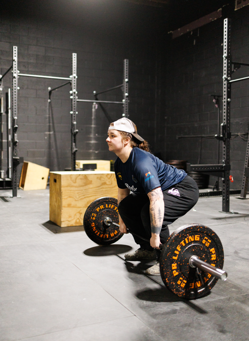
If you've been a member at Root Strength for any amount of time, you already know that physical therapy is part of what we do here. Our coaches hold Doctorates in Physical Therapy. Our programming is built around movement quality and long-term health. Injury prevention has always been woven into how we train.
What's changed is capacity. Our physical therapy department — Root Physical Therapy — is now fully operational and actively taking on new patients. The PT room is staffed, the schedule is open, and if you've been dealing with something you've been putting off, this is your sign to come in.
This isn't a referral to somewhere else. It's your own team, in your own gym, ready to take care of you.
The biggest gap in most athletic rehab isn't the quality of the PT — it's the disconnect between PT and the training environment. At Root Strength, those two things have always been the same program. Now we have the capacity to take on more of you.
What Root Physical Therapy Actually Is
Root Physical Therapy is our in-house PT department — not a separate business, not a clinic down the street. It's part of Root Strength, staffed by the same Doctors of Physical Therapy who coach your classes, design your programs, and understand your training inside and out.
That distinction matters. When your PT is also your coach — or works directly alongside them — there's no translation layer between what happens in a session and what happens on the gym floor. Your treatment plan isn't built around a generic athletic profile. It's built around you specifically: the weights you lift, the movements you do, and the goals you're working toward.
How It Works When PT and Coaching Are the Same Program
The standard rehab experience goes like this: you get injured, you go to an outside PT clinic, you do your exercises, you get discharged, and then you try to figure out on your own when it's safe to go back to training. That gap — between discharge and full return — is where most re-injuries happen.
At Root Strength, that gap doesn't exist. Here's what the process looks like when you come in as a PT patient:
-
01Assessment and coordinationYour PT evaluates what's happening and immediately coordinates with your coaches — what's safe, what isn't, and what modifications make sense right now. Everyone is aligned from day one.
-
02Modified training continues where possibleBecause your PT and your coaches are working from the same information, we can often keep you training — with adjustments — throughout your recovery. You stay active, maintain your conditioning, and don't lose the progress you've built.
-
03Progressive return to full trainingAs you recover, PT and coaching work together to gradually reintroduce full training. A clear, coordinated plan — not a generic discharge note and a wish of good luck.
-
04Back at full capacity — and staying thereYou return stronger and better informed about your body. And because your PT team is in the building with you, long-term support doesn't stop at discharge.
Insurance We Accept
We verify your benefits before your first appointment so there are no surprises. Most members end up paying little to nothing out of pocket.
Don't see your insurance listed? Reach out — we may still be able to help, or arrange a cash-pay rate that works for you.
Your Physical Therapy Team
Our PT department is staffed by four providers — all on-site, all part of the Root Strength team, each bringing a different clinical specialty.




Who Should Book an Appointment
You've been managing something for a while
A lot of members train through nagging injuries — the shoulder that flares on overhead press, the knee that complains after heavy leg days. If you've been tolerating something instead of resolving it, a proper assessment can identify what's actually going on and build a plan that fixes it.
You're recovering from something acute
A sprain, a strain, something that happened in training or outside the gym. The faster you start proper rehab, the faster you return to full training. And because your PT is in the same building as your coaches, that return is coordinated — not guessed at.
You want to stay ahead of injury
You don't need to be injured to work with our PT team. Movement screening, mobility work, and proactive care are some of the most valuable things you can invest in as an athlete who trains hard. If you're serious about longevity, this is part of the program.